Last Updated on June 22, 2023 by
What Can Cause Hip and Back Pain
“Why would someone call a podiatrist if they have hip pain?”
“The pain experienced in the hips can be caused by the feet being misaligned”
“Why Hip Replacements Don’t Always Get Rid of the Pain”
Below is the Journey of one of our clients who had both of her hip joints replaced, but still suffered from a considerable amount of hip pain.
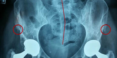
X-rays of her hips, see figure 14.6 below, showed:
- the hip replacements
- misalignment of the hips, with the left hip sitting higher than the right
- misalignment of the spine, as shown by the red line, the spine should be straight but in the x-ray it is clear this is not the case
- spurs on the hip bones, highlighted in red circles
“After two hip replacements this lady was still in pain”
Just looking at the x-ray of the hips it is clear there are a number of problems. The hip did not end up looking like this overnight, or in isolation. This amount of degeneration of the joints, and development of spurs, was happening for a protracted period of time before the hip surgery.
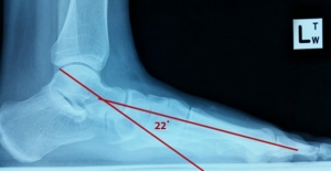
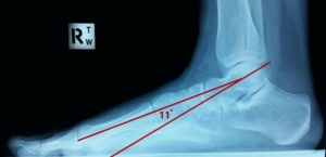
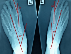
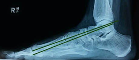
When I met this client our practitioner could see a number of subluxations in the joints of the feet, knees and hips just by looking at them. On account of the misalignment of the feet, there was years of uneven wear and tear in the knees and hips. Figures 14.7 to 14.9 below, are the x-rays of the feet showing subluxation in a number of the joints.
“After one of our practitioners took x-rays of the women’s feet the real issues could be seen”
The x-rays show:
- the left and right foot have significantly different angles
- the talus has dropped on both feet in the lateral x-ray, left worse than right
- there are bunions (1st toe misaligned) on both feet, right worse than left
- there are spurs on the hip bones
- there is severe arthritis of the hips
- both hip joints have been replaced
- the lower spine is curved
Overall it is clear to see there are a number of problems and these problems have been going on for a long time. Due to the subluxations in the joints of the feet there has been inefficient use of the muscles which in turn has led to uneven wear and tear in the hips. When the feet are out of alignment, then the body does not have a strong foundation to stand and walk on. This causes the legs, knees and hips to compensate, to rotate and to wear unevenly.
It is this misalignment which has led to years of wear and tear and eventually to the need for the hip replacements. Even after the replacements there was still pain and restrictions due to the fact that the underlying cause of the problems had not been addressed.
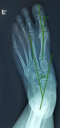
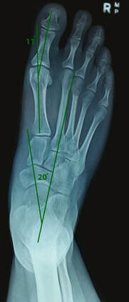
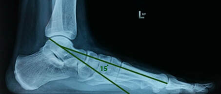
In this situation it was necessary to realign the joints of the feet, knees and hips in order to get the muscles to work efficiently and stop the grinding in the hip joint. Figures 14.10 to 14.13 show the new positions of the joints after three months of treatment.
“After only three months of treatment there was a dramatic difference in the x-rays and the women’s life”
On the DP x-ray the midline of the talus should form an angle of 15° with the midline of the foot. On the lateral x-ray the midline of the talus should form an angle of 0° with the midline of the foot.
Prior to starting treatment the DP x-ray showed the talus angle was 30° on the left and 20° on the right. After treatment the DP x-rays showed the talus angle was 23° on the left and 20° on the right.
Prior to starting treatment the lateral x-ray showed the talus angle was 22° drop on the left and 11° drop on the right. After treatment the lateral x-ray showed the talus angle was 15° drop on the left and 1° drop on the right.
This was a considerable improvement on the initial x-rays, overall there was 24° of change. On account of the feet being much better aligned the muscles in the legs were working more efficiently and the hip was not grinding unevenly. This client is now back to carrying out a number of high-impact activities, including looking after a very active six year old girl.
Any questions about hip pain? Just fill out the form below:
Or to see on of our practitioners please click the book in now button.

